What is blepharitis?

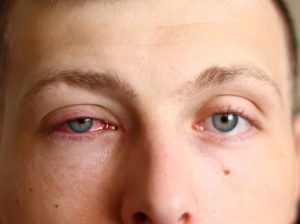
Blepharitis is an inflammation of the eyelids, often affecting both eyes along the edges of the eyelids. It typically occurs when the small oil glands near the base of the eyelashes become blocked, leading to irritation and redness. Various diseases and conditions can contribute to blepharitis, which is often a chronic and challenging-to-treat condition. While uncomfortable and unsightly, blepharitis usually does not cause permanent damage to eyesight and is not contagious

Symptoms of blepharitis include watery or red eyes, a gritty or burning sensation, greasy eyelids, itching, swelling, flaking skin around the eyes, crusted eyelashes, eyelid sticking, increased blinking, sensitivity to light, and occasional blurred vision that improves with blinking.

What is BlephEx?

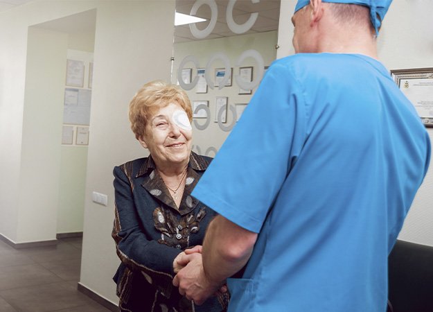
BlephEx is the first and only clinical treatment for blepharitis. Over 30% of patients experience symptoms of blepharitis.

BlephEx is an innovative, in-office procedure that allows clinicians to actively treat blepharitis. By reducing scurf and bacterial debris, which are the main causes of inflammatory lid disease, BlephEx improves the overall health of the eyelid and provides relief from chronic and irritating blepharitis symptoms.

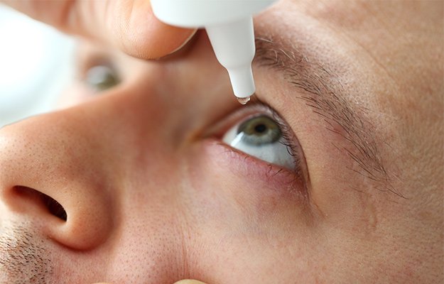
The BlephEx treatment is well tolerated and only takes a few minutes to perform, offering patients relief from the discomfort associated with blepharitis. Usually, several treatments are necessary. Additionally, BlephEx can lead to significant cost savings for patients by reducing their reliance on prescription drops and artificial tears.

How does it work?

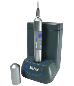
BlephEx is a painless, in-office procedure performed by eye care professionals. It utilizes a revolutionary patented BlephEx handpiece, which carefully and precisely spins a medical-grade micro-sponge along the edge of the eyelids and lashes, effectively removing scurf and debris while exfoliating the eyelids.

The patented micro-sponge is disposable, ensuring hygiene by using a clean one for each individual eyelid. Following the procedure, the eyes are rinsed thoroughly. Most patients find the procedure well tolerated, experiencing only a tickling sensation. Numbing drops are typically applied before treatment for added comfort.

After the procedure, patients receive instructions on maintaining clean eyelids with regular nightly lid hygiene. Since home treatments are only semi-effective, the procedure is typically repeated at 4-6 month intervals.

Why Choose BlephEx?

BlephEx offers a revolutionary solution for effectively cleaning eyelids, providing relief from blepharitis symptoms. It is the first and only clinical treatment for blepharitis, offering patients improved eyelid health and comfort.

While BlephEx is primarily designed to treat blepharitis by removing biofilm, debris, and scurf from the eyelids and eyelashes, it can indirectly help in the treatment of dry eye syndrome.

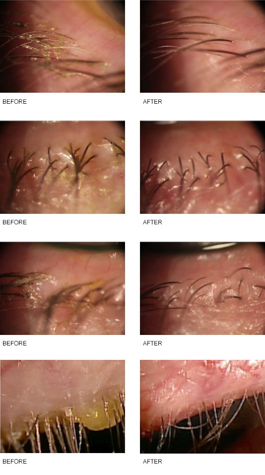
BlephEx Treatment before and after

Displayed below are images illustrating the contrasts in eyes affected by blepharitis before and after undergoing BlephEx treatment. Take a moment to observe and appreciate the remarkable effectiveness of this blepharitis treatment!

How does BlephEx help in dry eye treatment?

Dry eye syndrome often occurs alongside blepharitis, as the inflammation of the eyelids can disrupt the normal function of the meibomian glands, which are responsible for producing the oily layer of the tear film. This oily layer helps prevent the evaporation of tears and maintains the stability of the tear film, thus reducing the risk of dry eye symptoms.

By effectively cleaning the eyelid margins and improving eyelid hygiene, BlephEx can help restore the normal function of the meibomian glands and promote the secretion of healthy oils. This, in turn, can help stabilize the tear film, reduce tear evaporation, and alleviate dry eye symptoms such as dryness, irritation, and discomfort.

Additionally, by reducing inflammation and removing irritants from the eyelids, BlephEx can create a more favorable environment for tear production and distribution, further contributing to the management of dry eye syndrome.

It’s important to note that while BlephEx can be beneficial in managing dry eye symptoms associated with blepharitis, it may not be sufficient as a standalone treatment for severe cases of dry eye syndrome. A comprehensive approach to dry eye management may involve additional interventions such as artificial tears, prescription medications, lifestyle modifications, and in-office procedures like meibomian gland expression or thermal pulsation therapy, depending on the severity and underlying causes of the condition.

What is Pink Eye (Conjunctivitis)?

pink eye image

Pink eye, also known as conjunctivitis, is the inflammation of the transparent membrane that lines the eyelid and eyeball, called the conjunctiva. When the tiny blood vessels in this membrane become swollen and irritated, they become more visible, causing the whites of the eyes to appear pink or red. This condition can result from various causes, including viral and bacterial infections, allergies, or even a blocked tear duct in babies. Although pink eye can be bothersome, it rarely impacts your vision, and effective treatments are available to alleviate its discomfort. Given its contagious nature, early diagnosis and preventive measures can help contain its spread.

Symptoms

Common symptoms of pink eye include:

  • Redness in one or both eyes.
  • Itchiness in one or both eyes.
  • A gritty sensation in one or both eyes.
  • Eye discharge that crusts overnight, potentially making it challenging to open your eyes in the morning.
  • Excessive tearing.
  • Light sensitivity, also known as photophobia.

When to Seek Medical Attention

While pink eye is usually a mild condition, there are serious eye issues that may result in eye redness, pain, foreign body sensation, blurred vision, and light sensitivity. If you experience these symptoms, it’s crucial to seek urgent medical care. For those who wear contact lenses, discontinuing lens use as soon as pink eye symptoms develop is advisable. If your symptoms don’t improve within 12 to 24 hours, schedule an appointment with your eye doctor to rule out a more severe eye infection related to contact lens use.

Causes of Pink Eye

Pink eye can be caused by various factors, including:

  • Viruses
  • Bacteria
  • Allergies
  • Chemical splashes in the eye
  • Foreign objects in the eye
  • In newborns, blocked tear ducts

Types of Pink Eye (Conjunctivitis)

Viral and Bacterial Conjunctivitis 

The majority of pink eye cases result from viral infections, particularly adenovirus, but can also be triggered by other viruses such as herpes simplex virus or varicella-zoster virus. Bacterial conjunctivitis can occur alongside cold or respiratory infection symptoms, like a sore throat. Bacterial conjunctivitis can also result from improper contact lens hygiene.

Allergic Conjunctivitis

Allergic conjunctivitis affects both eyes and results from exposure to allergens such as pollen. This exposure triggers the release of inflammatory substances, including histamines, causing symptoms like red or pink eyes, intense itching, tearing, and nasal discharge. Allergic conjunctivitis is not contagious.

Conjunctivitis Resulting from Irritation

Irritants such as chemical splashes or foreign objects in the eye can lead to conjunctivitis. Flushing and cleansing the eye can sometimes cause redness and irritation. Most irritation-related symptoms, including watery eyes and mucous discharge, resolve within a day. However, if symptoms persist or are caused by caustic chemicals, immediate medical attention is essential to prevent potential eye damage.

Risk Factors for Pink Eye

Factors that increase the risk of developing pink eye include exposure to someone with viral or bacterial conjunctivitis, exposure to allergens for allergic conjunctivitis, and the use of contact lenses, especially extended-wear lenses.

pink eye image 2

Possible Complications

In both children and adults, pink eye can lead to corneal inflammation, affecting vision. Timely evaluation and treatment by a healthcare provider can reduce the risk of complications. Seek medical attention if you experience eye pain, sensation of a foreign object in your eye, blurred vision, or light sensitivity.

Preventing The Spread Of Pink Eye

To prevent the spread of pink eye, practice good hygiene, including:

  • Avoid touching your eyes with your hands.
  • Wash your hands frequently.
  • Use clean towels and washcloths daily.
  • Do not share towels or washcloths.
  • Change pillowcases regularly.
  • Dispose of old eye cosmetics, such as mascara.
  • Do not share eye cosmetics or personal eye care items.

Pink eye is no more contagious than the common cold. If good hygiene practices are followed, individuals can typically return to work, school, or child care once their symptoms clear up. However, in situations where close contact with others is involved, it may be advisable to stay home until symptoms have subsided.

Preventing Conjunctivitis in Infants

Newborns are particularly susceptible to bacteria present in the mother’s birth canal. These bacteria, often asymptomatic in the mother, can lead to ophthalmia neonatorum, a severe form of conjunctivitis in infants. To prevent this, antibiotic ointment is applied to newborns’ eyes shortly after birth.

Is it possible to contract pink eye by making eye contact with an infected person?

Pink eye, or conjunctivitis, is typically spread through direct or indirect contact with the eye secretions of an infected person. Simply looking at each other’s eyes without any direct eye secretion contact is unlikely to spread pink eye. However, close contact, such as being in the same room or using the same items that have come into contact with infected eye secretions (like towels or makeup), can increase the risk of transmission. It’s essential to practice good hygiene and avoid sharing personal items to reduce the risk of spreading pink eye. If you suspect you or someone you know has pink eye, it’s a good idea to take precautions to prevent its spread.

Which eye conditions can be mistaken for pink eye?

  • Episcleritis: This is an inflammation of the episclera, a clear layer covering the sclera (the white part of the eye). It can cause redness and mild discomfort, resembling pink eye.
  • Keratitis: Inflammation of the cornea can lead to eye redness, pain, and tearing, which might be confused with pink eye.
  • Iritis/Uveitis: These conditions involve inflammation of the iris and uvea, respectively, and can cause eye redness, pain, and light sensitivity.

It’s essential to consult with an eye care professional for an accurate diagnosis if you’re experiencing eye symptoms similar to pink eye, as they can differentiate between these conditions and recommend the appropriate treatment.

Treatments for Pink Eye

Regardless of the type of pink eye, you can help your recovery by practicing good eye hygiene, avoiding sharing personal items like towels or makeup, and refraining from wearing contact lenses until your symptoms have resolved. If symptoms persist worsen, or if you have severe eye pain, it’s essential to seek medical attention for proper evaluation and treatment.

Are you suffering from pink eye? Don’t delay, seek relief and expert care at the European Eye Center. Our dedicated team is here to provide the best treatment and guidance for your eye health. Contact us now to schedule your consultation and start your journey to brighter, healthier eyes today!

Glaucoma is the leading cause of irreversible blindness in adults aged 40 and above. Keep reading to discover the basics of glaucoma and its first signs.

What Is Glaucoma?

Glaucoma is a progressive eye disease that damages the optic nerve inside the eye caused by increased internal eye pressure.

eye-disease-Symptoms-as-body - What are the Early Sign of Glaucoma?

When left untreated, it will cause permanent vision loss and lead to blindness. The problem with glaucoma is, it can go undetected for years with few or no warning signs, but in the mean time causes irreversible damage to the optic nerve, the link between your eye and brain.

It is estimated that approximately 64 million people have glaucoma worldwide. While anyone can develop glaucoma due to various external factors, the condition is the most prevalent among adults over the age of 40.

Glaucoma tends to run in families. If you have glaucoma in your family, you should do a glaucoma screening yearly.

Causes

Your eyes are filled with aqueous humor. This transparent, water-like substance provides nutrition to the eye and maintains the pressurized shape of the eye. It’s continuously produced and drained from the eye. Patients with glaucoma don’t drain excess fluid properly and pressure builds up inside the eye, causing damage to the optic nerve.

Usually, this is a slow, insidious process (open-angle glaucoma) but in some cases, an acute rise in eye pressure due to closure of the drainage, causes acute or closed-angle glaucoma. This is a medical emergency.

Other possible causes of glaucoma include blunt or chemical trauma, severe eye infections, blood vessel blockage inside the eye, and inflammatory conditions. Despite being unlikely, eye surgery may give rise to glaucoma.

Risk Factors

Glaucoma usually affects adults over the age of 40, but also young adults, children and even infants can have glaucoma. Risk factors include:

  • Being age 40 and over
  • Having family with a history of glaucoma
  • Being of Hispanic, Asian, or Black descent
  • Having diabetes
  • Having Sickle Cell Anemia
  • Having hypertension (high blood pressure)
  • Having previous eye injury or surgery
  • Having thinner corneas
  • Taking certain corticosteroid medication
  • Having very dense cataract
  • Having elevated eye pressure

Early Signs & Symptoms of Glaucoma

Most people with (open-angle) glaucoma don’t have any symptoms. If symptoms appear, it’s usually late in the disease and the main sign is loss of peripheral vision. Because this happens so slowly over time, people are usually unaware of it.

Sometimes people can develop acute or closed-angle glaucoma. In this case, symptoms and damage develop quickly. This is an eye care emergency.

You should seek help immediately if you develop the following symptoms:

  • Intense eye pain or headache
  • Eye redness
  • Halos, rings, or rainbows around lights
  • Vision Loss
  • Nausea and vomiting
  • Eye that looks hazy

Diagnosis & Treatment

Glaucoma treatments

How to Diagnose Glaucoma

It generally doesn’t take long to check for glaucoma. The examination is quick and includes a vision check, tonometry (measurement of eye pressure) and after dilation of your pupils with eyedrops, a visual check of your optic nerve. When glaucoma is suspected, special imaging to document the state of the optic nerve and a visual field test to check for peripheral vision loss will also be performed.

How to Treat Glaucoma

Glaucoma (open-angle) is usually treated with medication but sometimes requires surgery.

Some of the standard treatments for glaucoma include:

  • Eye drops — to lower eye pressure.
  • Oral medications — beta-blocker or carbonic anhydrase inhibitors slow fluid production and improve drainage.
  • Surgery — trabeculectomy helps drain fluid through a newly created channel. Other procedures include iridotomy and cyclophotocoagulation.
  • Cataract surgery

Acute (closed-angle) glaucoma is a medical emergency and is treated with a laser procedure.

Glaucoma is a lifelong disease and requires continuous follow-up.

If you’re over 40,  have glaucoma in your family or experience symptoms of glaucoma, contact EEC today for an eye exam!

Every patient deserves top-quality eye care from us. European Eye Center focuses on providing Western-standard services and determining appropriate treatment plans to help patients restore their vision in a feel-like-home atmosphere.

We are happy to help if you have questions about the first signs of glaucoma or any other vision conditions. Don’t hesitate to connect with us at info@europeaneyecenter.com.

Contact us today to schedule an eye care service!

In this article, we will learn about a macular hole, including its definition, symptoms, causes, and treatment.

What Is a Macular Hole?

macular-pucker-and-retinal-detachment-in-woman-Symptoms-as-body

Before talking about macular holes, let’s first define what the macula is. The macula is the part of the retina that is essential for central vision and color vision. The central vision allows you to focus on details.

A macular hole is literally a hole that develops on the macula, due to the separation of nervous cells within the retina. Over time this hole can grow in size and cause more central vision loss.

Despite its significant impact on vision (it may lead to permanent vision loss), a macular hole is relatively rare, with only 8 out of 100,000 people being affected with this condition throughout their lifetime. Most macular holes develop with age (age 55 and above) and women are at higher risk of developing macular holes than men. If you develop a macular hole in one eye, you have a 10-15% chance of developing one in the other eye.

Symptoms

Macular holes develop slowly. You may start to notice a slight blurriness or distortion when looking at objects.

Possible symptoms may include:

  • Decreased sharpness of vision, regardless of looking far or near.
  • Distorted or blurred vision when looking straight ahead.
  • Straight lines or objects become bent or wavy.
  • Difficulty reading, writing or driving.
  • Blurred or dark spots in the middle of the visual field.
  • Inability to see a person’s nose or eyes.
  • Severe changes in visions that feel like looking through frosted glass.

Causes

Aging is the leading cause of macular holes. Other causes include:

  • Eye injury or blunt trauma
  • Complicated myopia
  • Diabetic eye disease

Diagnosis

An OCT, short for Optical Coherence Tomography, is used to diagnose macular holes. It’s a quick and painless imaging test, that scans the retina allowing to assess the macula and retina with high-definition pictures. It can also rule out other eye conditions with similar symptoms.

Treatment

A tiny, insignificant macular hole may occasionally resolve on its own, but it will have to be monitored closely.

Most macular holes require surgery, and the most common procedure is a vitrectomy. During the procedure, the vitreous gel is replaced with a gas and air mixture. This bubble serves as a temporary bandage that holds the macula in place till it’s healed.

Following the surgery, you may have to cut back on some activities, such as flying or diving, lifting, or bending.

If you experience any of the symptoms of a macular hole, contact EEC today for an eye exam!

Every patient deserves top-quality eye care from us. European Eye Center focuses on providing Western-standard services and determining appropriate treatment plans to help patients restore their vision in a feel-like-home atmosphere.

We are happy to help if you have questions about macular holes or any other vision conditions. Don’t hesitate to connect with us at info@europeaneyecenter.com.

Contact us today to schedule an eye care service!

In recent years, many people have expressed concerns about blue light and whether it contributes to various eye issues. Is blue light bad for your eyes? Let’s go over the basics of blue light, health benefits, possible effects on our eyes, and the use of blue-light-blocking glasses.

Woman-watching-TV-at-home-and-holding-the-remote-control-What-Is-Blue-Light-as-body

What Is Blue Light?

Blue light is a portion of the visible light spectrum and it’s all around us. The sun is the biggest source of natural blue light. Nowadays we are more exposed to blue light because of our widespread use of devices that use LED technology, like computer screens and smartphones. This LED technology emits high amounts of blue light. The concern about blue light finds its origin in its high energy and proximity to UV light in the electromagnetic spectrum.

UV light, which is invisible, has long been linked to eye and skin damage. Thankfully there is very little indication that blue light emitted from LED screens is harmful. The LED screens are simply not bright enough. The natural blue light from the sun far exceeds the amount emitted from a screen.

The Health Benefits of Blue Light

boy-lies-with-toy-bear-in-bed-under-a-blanket-How-Blue-Light-May-Help-with-Health-as-body

Blue light, mostly emitted by the sun, has been proven to promote alertness, boost mood, improve memory and cognitive function, enhance healthy sleep by maintaining a wel-lregulated circadian rhythm, and help with some skin conditions.

Is Blue Light Bad For Your Eyes?

guy-massaging-the-nasal-bridge-after-working-on-a-laptop-Is-Blue-Light-Bad-For-Your-Eyes-as-body

Our eyes are built in a way that provides protection against some kinds of light. The cornea and lens protect the retina, at the back of the eyes, against harmful UV rays. They are not good at blocking blue light from entering the eyes. This has sparked the debate about whether extended use of electronic devices can give rise to eye disorders. Thankfully there is very little indication that blue light is responsible for any eye pathology, whether it’s from the sun or from an LED screen which is far less bright.

Extended use of LED screens is linked to digital eye strain, but this is mostly due to the fact that we blink far less often than normal when looking at a screen, thus causing our eyes to dry out. So it’s not the blue light, but the fact that we blink less often that causes eye problems.

Symptoms of digital eye strain include:

  • Eye irritation or soreness
  • Tired eyes
  • Blurred vision
  • Headaches or fatigues
  • Tearing

Do Blue-Light-Blocking Glasses make a difference?

woman-going-through-paperwork-How-to-Reduce-Blue-Light-Exposure-as-body

Blue-light-blocking glasses will not make a big difference during the day.
Besides there being very little evidence blue light emitted from LED devices is harmful for your eyes, most blue-light-blocking glasses will only filter 5-40% of blue light anyway.

Wearing them at night, might be useful, since blue light inhibits the release of melatonin, so when exposed in the evening, blue light might cause difficulties to fall asleep.

If you have questions about blue light or suffer from digital eye strain symptoms, contact EEC today for an eye exam!

Every patient deserves top-quality eye care from us. European Eye Center focuses on providing Western-standard services and determining appropriate treatment plans to help patients restore their vision in a feel-like-home atmosphere.

We are happy to help if you have questions about blue light or any other vision conditions. Don’t hesitate to connect with us at info@europeaneyecenter.com.

Contact us today to schedule an eye care service!

Aging is the leading cause of developing a cataract. Your vision becomes blurry as a result. Sometimes, cataract formation can also be the result of taking particular drugs, illnesses, or trauma. If you think your sight has become blurry or your color vision has become less bright, you may have developed cataracts.

EEC-Cataract concept. Senior woman_s eye-as-What Is Cataract Surgery_

Please read this post to thoroughly understand cataract surgery and how to prepare for it.

What Is Cataract Surgery?

Your eye’s natural lens is usually transparent. When a cataract develops, your lens becomes cloudy and this impairs your vision.

When ophthalmologists perform cataract surgery, they remove the lens of your eye and replace it with an artificial lens.

Cataract surgery is a safe and very common procedure. Most cataracts develop in adults as a result of aging.

When Do You Need Cataract Surgery?

EEC-Eye diseases age related-as-When Do You Need Cataract Surgery_

Cataracts will slowly make you see more blurry and colors will appear less bright. If a cataract starts interfering with your daily activities, it’s time to think about cataract surgery. But you don’t have to wait that long. Nowadays doctors perform cataract surgery much earlier than before.

If you have other eye conditions, like age-related macular degeneration or diabetic retinopathy, and your eye doctor’s ability to monitor or treat these conditions diminishes because of cataracts, he or she will definitely recommend doing cataract surgery.

Cataracts don’t impair your vision right away unless they are caused by trauma. They usually worsen over time. Slowly your prescription will start to change, you may find reading, especially in low light conditions, more challenging, or you may start noticing halos or glare around lights when driving at night. Colors will also appear less bright.

Little by little your vision will deteriorate and at some point, even a higher prescription will not help you anymore.

At that point, the only way to improve your vision is through cataract surgery, to remove the cataract (the cloudy lens) and insert an intraocular lens.

An ophthalmologist can easily confirm your cataracts with a slit lamp examination.

Besides diagnosing the cataract, It’s also very important to examine the rest of the eye, especially the retina. If there is a retina problem, it might have to be treated first, in order for the cataract surgery to be beneficial.

Doing cataract surgery as soon as symptoms start to appear, or waiting till it’s really necessary, is up to the patient unless there are other complications, that require intervention.

What to Expect

EEC-The operation on the eye-as-What to Expect

Before the Surgery

An extensive eye exam needs to be performed to determine the condition of the retina, the density of the cataract, and the size of the eye in order to choose the right implant lens.

During the Surgery

Cataract surgery is usually performed as an outpatient procedure, which usually takes 30 to 1 hour and allows you to recover at home.

When you arrive at the hospital:

  • The nurse will dilate your pupil by putting eyedrops in your eye.
  • Local anesthetics, in the form of eye drops, will be used to numb the eye. Your surgeon might give you a sedative to relax when it’s deemed necessary.
  • Through a tiny hole, the cataract is sucked out of the eye after being emulsified, and through the same hole, an artificial lens is inserted. The lens capsule at the back of your eye is kept whole so the artificial lens can be placed there. The procedure is called: Phaco-emulsification. This is practically the only procedure used nowadays to remove cataracts.

After the Surgery

Immediately after the procedure, your eye will be covered with an eyepatch. This will be removed the next day at your first post-op consultation. Initially, your vision might still be a little blurry, but this is normal. Some itching and discomfort will also be present during the first few days. Be careful not to rub or squeeze your eye. Your doctor might instruct you to wear a protective shield. Eye drops will have to be used for a few days after the surgery.

Your doctor will schedule a few follow-up appointments after the surgery.

The day after the surgery, when the eyepatch is removed, you will see an improvement in your vision. Sometimes, due to swelling, it can still be a little blurry, but that will clear up in a few days.

If you have any eye problems or think you have cataracts, schedule an exam today to find the right diagnosis and best treatment.

Every patient deserves top-quality eye care from us. European Eye Center focuses on providing Western-standard services and determining appropriate treatment plans to help patients restore their vision in a feel-like-home atmosphere.

We are happy to help if you have questions about cataract surgery. Don’t hesitate to connect with us at info@europeaneyecenter.com.

Contact us today to schedule an eye care service!

In this article, we will learn about myopia, a common vision condition affecting billions of people worldwide, including causes, symptoms, complications, and treatments.

Teenager-squinting-her-eyes-while-using-laptop-Symptoms-as-body

What Is Myopia?

If you have problems seeing objects or items from a far distance or you need to move closer to see them clearly, you probably have myopia or nearsightedness.

Myopia is a common refractive error condition. It’s been estimated that by the year 2050, about 5 billion people worldwide will be affected by it. The prevalence of myopia is particularly high in Asian countries:  80-90% of young Asian people are nearsighted.

Progressive myopia means the correction to see clearly increases by more than 0.5D per year. This form of myopia typically affects children and teens.

Causes

Usually, myopia occurs when the eyeball is too long. In a person with normal vision, the light rays enter through the pupil and are focused directly on the retina, giving a clear image. When the eyeball is too long, the light rays are focused in front of the retina, giving a blurry image.

In case of progressive myopia, the eyeball grows longer and longer as the child continues to grow, increasing the refractive error.

There is definitely a genetic component of myopia; children of parents with myopia have a much higher risk of having or developing myopia.

People of Asian descent have a higher risk of myopia.

An excessive amount of close-up tasks like reading, computer work or watching videos on one’s phone is linked to progressive myopia.

Recent studies show that children who don’t spend enough time outdoors (1.5 h per day) are also more likely to develop myopia.

Symptoms

A person or child may have myopia if they have the following symptoms:

  • Blurred vision and need to get closer to see clearly
  • Unaware of distance items or objects
  • Excessively blinking
  • Headaches or fatigue
  • Persistent squinting
  • Eyestrain
  • Rubbing eyes frequently

Adults with myopia may experience vision difficulties when driving.

Children with myopia may complain about not being able to read or see what is written on the blackboard at school and feel like they need to sit closer to watch TV or lower their heads while writing.

Complications

What are the long-term effects of myopia other than the need to wear eyeglasses or contacts every day?

Health experts have pointed out that people with high myopia have increased risks of the following eye diseases:

  • Cataracts
  • Glaucoma
  • Retinal detachment
  • Macular degeneration
  • Myopic maculopathy.

These above eye conditions can cause permanent vision loss if no action is taken. Therefore, regular eye exams are highly recommended to manage myopia and detect early signs of sight-threatening diseases.

Diagnosis

The first step in diagnosing myopia is a visual acuity test. Usually, this is done with a Snellen Chart. Your eye doctor will ask you to read the lines of letters decreasing in size till you can no longer read them.

Children may receive eye drops to eliminate accommodation and to objectively measure their visual acuity with the Snellen Chart

In case of progressive myopia, a regular biometry (ultrasound) will be performed to follow the increase in eyeball length.

Treatments

The most common treatment for myopia is a prescription for eyeglasses or contact lenses.

If you wish to get rid of myopia more permanently, refractive surgery such as LASIK, SMILE, or PRELEX is another option. These procedures are only recommended for adults with myopia that is no longer progressing. Otherwise, nearsightedness may make its way back, and you will need to wear glasses again.

During refractive surgery, eye surgeons will either replace your natural lens or use a laser to reshape your cornea, both allowing light to focus precisely on the retina.

An innovative and safe technique that has been developed and proven to help people with myopia — even children — see more clearly is Ortho-K (orthokeratology). Ortho-K corrects vision by using a rigid contact lens to temporarily reshape the curved cornea — similar to what dental braces usually do. The lenses are worn at night and removed in the morning, so you have clear vision during the rest of the day.  Ortho-K does not correct myopia permanently. It only takes away the need to wear glasses or contacts during the day.

Treatment with daily Atropine drops can slow down the progression of myopia in children..

Prevention

Myopia can not be entirely prevented, but there are some good eye health habits to prevent progression and protect your eyes in general, like:

  • Have regular eye exams
  • Read or do near-focused tasks with good lighting and at a distance equal to the length of one’s forearm.
  • Watch TV from at least 3m distance
  • Outdoor activities for at least 1.5h a day
  • Wear protective eyewear when playing outdoor sports
  • Wear sunglasses to limit sun exposure
  • Wear corrective lenses as directed by your doctor
  • Rest your eyes with the 20/20/20 rule after using digital devices for a long time
  • Adapt a balanced and healthy diet
  • Quit smoking
  • Exercise
  • Keep your chronic conditions under control (diabetes, hypertension, and cholesterol levels)

If you experience blurry vision or eye strain, contact EEC today for an eye exam!

Every patient deserves top-quality eye care from us. European Eye Center focuses on providing Western-standard services and determining appropriate treatment plans to help patients restore their vision in a feel-like-home atmosphere.

We are happy to help if you have questions about myopia or other vision conditions. Don’t hesitate to connect with us at info@europeaneyecenter.com.

Contact us today to schedule an eye care service!

Have you ever looked someone in the eye and noticed they have 2 different colored eyes? This is called heterochromia and is usually of no further concern.

What is Heterochromia?

What is Heterochromia?

When a person’s irises are different in color, they have heterochromia.

There are a few kinds of heterochromia. When the complete iris is a different color from the other, it’s called complete heterochromia. When only a part of the iris has a different color, it’s called partial heterochromia. Central heterochromia means there is only an inner ring on the iris with a different color.

Causes of Heterochromia

The color of the irises is determined by the amount and distribution of a pigment called melanin. Heterochromia is caused by either lack or excess of melanin in one eye.

Most cases are hereditary, but sometimes heterochromia can be caused by certain diseases or an injury that causes one eye to change color.

An infant can be born with heterochromia or develop it soon after birth. This is called congenital heterochromia. In most cases, this is of no concern, and there will be no other symptoms or health issues.

However, in some cases, there can be underlying conditions. An ophthalmologist should therefore examine a newborn or infant with heterochromia to rule out any other health issues.

When a person develops heterochromia later in life, it’s called acquired heterochromia and is caused by disease or injury.

Causes can be:

  • Eye injury or foreign object in the eye
  • Eye surgery
  • Bleeding or inflammation in one eye
  • Glaucoma and some medications to treat it
  • Tumors of the iris
  • Diabetes
  • Central retinal vein occlusion
  • Neurofibromatosis

When an adult develops heterochromia later in life, they should be examined by an ophthalmologist to determine the cause. Treatment will be focused on the underlying condition. If no other eye issues exist, no treatment is needed for the heterochromia.

Some famous people with heterochromia include:

  • Keifer Sutherland
  • Kate Bosworth
  • Demi Moore
  • Dan Akroyd
  • David Bowie developed heterochromia due to a childhood injury.

Every patient deserves top-quality eye care from us. European Eye Center focuses on providing Western-standard services and determining appropriate treatment plans to help patients restore their vision in a feel-like-home atmosphere.

We are happy to help if you have questions about the heterochromia. Don’t hesitate to connect with us at info@europeaneyecenter.com.

Contact us today to schedule an eye care service!

In this article, we will learn more about 20/20 vision, a standard ratio to express a person’s visual acuity, its definition, ways to correct less than 20/20 vision, and tips for keeping your eyes healthy.

20/20 vision

What does 20/20 Vision mean?

The term 20/20 vision expresses your level of visual acuity or ability to see sharp from a specific distance (20 feet), as determined through an eye exam. In the UK ophthalmologists sometimes still use the ratio 6/6 (20 feet = 6 meters).

Visual acuity is an expression that describes how sharp or clear you can see from a specific distance.  20/20 vision is considered normal, meaning the average person can see the chart sharp from 20 feet. If you have 20/100 vision, you can see at 20 feet what the average person can see at 100 feet.

Is 20/20 Vision Common?

In the US, it is estimated that 1 in 3 adults has 20/20 vision. In Asia, this ratio is lower.

Can Vision Be Better Than 20/20?

Yes, your vision can be better than 20/20. Having 20/20 vision means your vision is average or normal, but some people have 20/15 or 20/10 vision, meaning they can see objects sharp at 20 feet that ordinary people see sharp at 15 or 10 feet away.

When your vision is less than 20/20, for example, 20/50, you can see objects sharp at 20 feet that the average person can see sharp at 50 feet away.

What Causes Less Than 20/20 Vision?

The most common reasons for less than 20/20 vision are refractive errors, such as nearsightedness, farsightedness, or astigmatism.

But specific eye or systemic diseases can also affect a person’s ability to see clearly, including:

  • Multiple sclerosis
  • Diabetes
  • Thyroid disease
  • Hypertension
  • Glaucoma
  • Cataracts
  • Macular degeneration
  • Strabismus

If your vision is less than 20/20 due to refractive errors, your eye doctor will prescribe a prescription for eyeglasses or contact lenses, or you can opt for vision corrective surgery.

Tests to Assess Visual Acuity

Man-putting-liquid-drops-in-his-eye-Tests-to-Assess-20-20-Vision-as-body

Visual acuity is typically tested with a standardized reading chart. The most commonly used is the Snellen chart, with eleven rows of letters decreasing in size. From 20 feet away, you are instructed to read the rows of letters until you no longer can.

Your visual acuity will be expressed as a fraction where the first number indicates the distance, and the second number refers to the letter size.

If you have a visual acuity better than average, your second number is smaller than 20 (i.e., 20/15). In contrast, if your second number is greater than 20 (i.e., 20/80), it signifies that your visual acuity is less than average.

Visual acuity and more…

Visual acuity is not everything, it’s just one part of your vision.

The complete visual function also includes depth perception, the field of vision, color vision, eye movement, and binocular vision. A comprehensive eye exam will include all these facets of your vision and overall eye health.

How to keep your eyes healthy

Your eyes, as the rest of your body, are susceptible to aging. Here are some tips to keep your eyes as healthy and your vision as sharp as possible:

  • Have regular eye exams
  • Quit smoking
  • Stay physically active
  • Maintain healthy weight
  • Control your cholesterol levels
  • Keep track of blood glucose levels if you have diabetes
  • Wear sunglasses and protective eyewear
  • Let your eyes rest after using digital devices for prolonged periods
  • Adopt a balanced and healthy diet with adequate nutrients and vitamins for eye health

Schedule an eye exam at EEC to check your visual acuity today.

Every patient deserves top-quality eye care from us. European Eye Center focuses on providing Western-standard services and determining appropriate treatment plans to help patients restore their vision in a feel-like-home atmosphere.

We are happy to help if you have questions about the 20/20 vision. Don’t hesitate to connect with us at info@europeaneyecenter.com.

Contact us today to schedule an eye care service!

When you go to the eye doctor to measure your eyesight to get new eyeglasses, you will be given a paper with numbers, letters, and symbols. This is your eyeglasses prescription.

Not everyone knows how to read these prescriptions properly. Today’s article will guide you to read your eyeglasses prescription.

Hand-holds-doctor-eyeglass-prescription-How-to-Read-an-Eyeglasses-Prescription-ss-feat

OD & OS: What does this mean?

  • OD represents the right eye. It is short for Oculus Dexter, Latin for right eye.
  • OS represents the left eye. It is short for Oculus Sinister, Latin for the left eye.
  • OU might sometimes be written in a column in your prescription. OU is short for Oculus Uterque, meaning both eyes in Latin.

These terms are not only standard in eyeglasses prescriptions but also for contact lenses and eye medicines. Some doctors prefer using RE (right eye) and LE (left eye) instead of OD and OS.

portrait-of-young-man-having-eye-astigmatism-myopia-OD-vs.-OS-What-are-they-ss-body

Sphere (SPH)

The term “sphere” indicates the amount of lens power measured in diopters (D) needed to correct nearsightedness or farsightedness. The term ‘sphere’ means the correction is ‘spherical’ or equal in all the meridians of the eye.

You are nearsighted if a (-) symbol comes with the number. If the number is preceded by a (+) sign, you are farsighted. So, for example, you see in the sphere section +5.00 — this means there are five diopters of farsightedness.

Number zero means your eyes need no correction. Thus, the further you are from zero, on either the minus or plus side, the higher your prescription is and the lower your uncorrected vision.

Cylinder (CYL)

The cylinder number indicates the power needed to correct astigmatism. If this box is empty or reads 0, astigmatism is not present or it’s so small it doesn’t need to be corrected. The cylinder power is always a minus number.

The term ‘cylinder’ means that this lens power is not spherical.

If you have astigmatism, your eye is shaped more like a rugby ball instead of a football, and this needs to be corrected in this way.

Axis

If your prescription has a cylinder power, it must be followed by an Axis number.  Axis indicates the location of astigmatism on the cornea. The axis, a number from 1 to 180, is the lens meridian that is 90 degrees away from the meridian that contains the cylinder power to correct astigmatism.

The number 90 refers to the vertical meridian of the eye, while 180 corresponds to the horizontal meridian.

Add

‘Add’ refers to ‘Addition’ or the added magnifying power applied to the bottom part of multifocal lenses to correct presbyopia. Presbyopia is a part of aging, indicating the gradual loss of your eye’s ability to focus on nearby objects.

The number appearing in the Add section of the prescription always goes with a “plus” power. It typically falls between +0.75 to +3.00 D and will be the same power for both eyes.

Prism

Prism is designed to correct vision for people with diplopia or double vision. These conditions make patients see two distinct images of one object. The prism helps align the two images so that only one image is visible.

Prism is not always present in eyeglasses prescriptions. When it is, the amount of prism is calculated in either metric or fractional English units.

The direction of the prism is indicated by noting the relative position of its “base.”

  • BU = base up
  • BD = base down
  • BI = base in
  • BO = base out

Example of Reading an Eye Prescription

SPH CYL Axis Add Prism
OD -5.00 +3.00 0.5 BD
OS -5.50 -1.00 180 +3.00 0.5 BU

OD = the right eye:

  • -5.00 D sphere for nearsightedness correction.
  • There are no numbers in the cylinder power or axis section, which means the patient doesn’t have astigmatism.

OS = the left eye:

  • -5.50 D sphere for nearsightedness correction.
  • -1.00 D cylinder for the astigmatism correction, with an axis at 180 degrees.

Both eyes:

  • Add section is +3.00 D for the correction of presbyopia, the ‘reading correction’
  • Prismatic correction of 0.5 prism diopter in each eye.

If you have eye disorders or need eyesight measurements, schedule an exam today to find the best treatment.

Every patient deserves top-quality eye care from us. European Eye Center focuses on providing Western-standard services and determining appropriate treatment plans to help patients restore their vision in a feel-like-home atmosphere.

We are happy to help if you have questions about eyeglasses prescriptions. Don’t hesitate to connect with us at info@europeaneyecenter.com.

Contact us today to schedule an eye care service!